Histology
Courtesy of Lab of Pathology Department on Medicine School of Ribeirão Preto – USP
01
Study
Group
N= 29 Wistar rats
Weight mean : 421g
Origin of animals : Biotério Central of Campus of Ribeirão Preto of São Paulo University
Maintenance : Experimental Pathology Lab of Pathology Department on Medicine School of Ribeirão Preto – USP
Animals keeping : polypropylen boxes
Animals food : basic diet of lab and water
Light conditions : natural light/dark cycles under controlled basic conditions
Experimentators : A.Tenenbaum,R.Mené,Kim

02
Animals Sacrifice & Histologic Samples
The mice were euthanized in CO2 chambers.
- Incision of the skin with a blade nº 23
- Finding and isolation of the pretibial muscle using Metzenbaum scissors
- Section of tendons with a Mayo scissors.
- Fixation of Portions of the muscles on sections of cork oak
- Embedding using 50% formaldehyde solution - 10% buffered and 50% of pure paraffin for conventional optical microscopic study.
- Staining Sections as routine histological process.
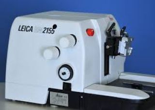
- Microtome used : Leika RM 2155
- Coloration of muscle slices with
-hematoxilin-eosin
-Masson´s trichrome
-and red picro-sirius.
- Histologic examination was realized with a conventional microscope (Olympus BH-2).
- Observation under polarized lampe
- Photographic documentation was realized with a Leika DMR microscope joined to a digital camera Leika CD 300F and compatible PC.
03
Material
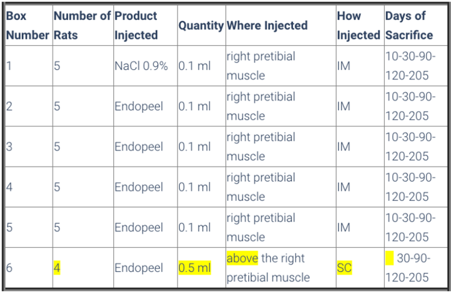
- Animals were divided in 6 boxes, so that boxes from number 1 to 5 had 5 rats and box number 6 had 4 rats
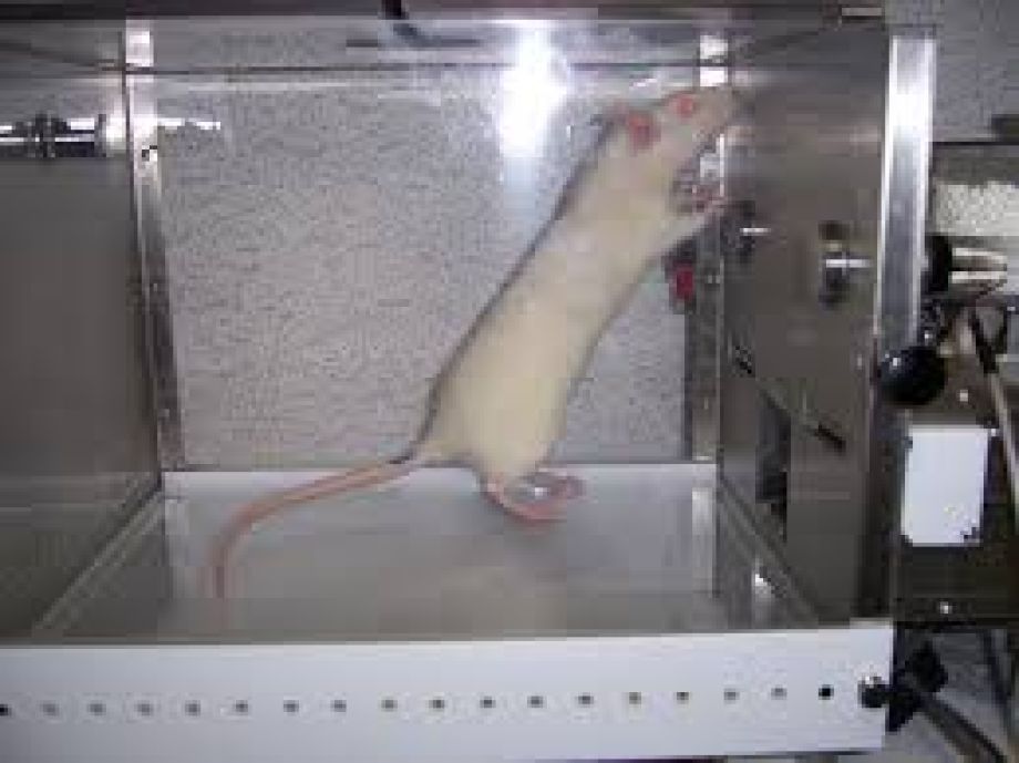
- All animals were submitted to a walking evaluation by footprint impression on special paper , and previous immersion of back legs on a soap solution.
- Box 1 had the control group, that received the application of 0,1 ml saline solution ( NaCl 0,9%) on the right pretibial muscle.
-
The mice of boxes 2 to 5 received 0,1 ml of Endopeels Original Main Product on the same muscle as control group.
-
Box 6 was constituted of 4 rats, and each one received 0,5 ml of Endopeels original main product injected on the subcutaneous layer.
-
All the animals were submitted to walking evaluation before and after their respective injections.
04
Methods
05
Control
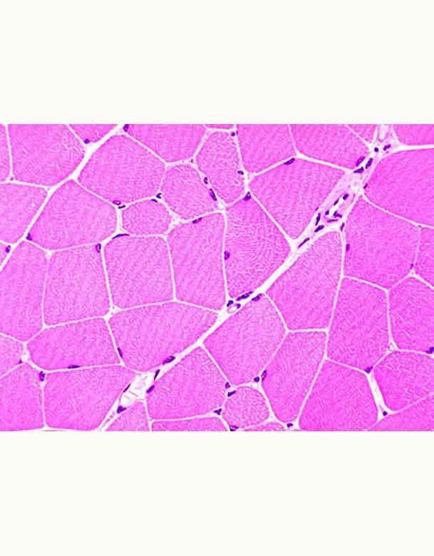
Comment : Nothing to declare after saline solution injection
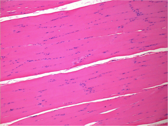
L:Pretibial-No treatment
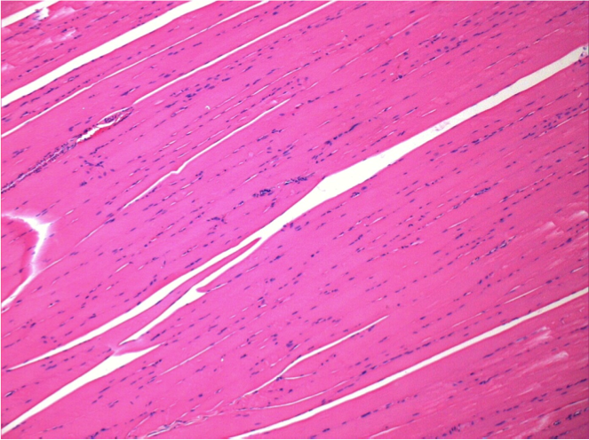
R:Pretibial-After 0.1 ml NaCl 0.9% IM
06
10 days after Endopeel Injection
10 days after Endopeel Injection 0.1ml in the right pretibial muscle.
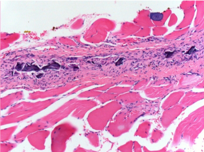
Here you may see the formation of the vacuoles which are surrounded by lymphocytes. Vacuoles are different from tissue necrosis . The presence of lymphocytes is related to the permeability of the cell membranes.
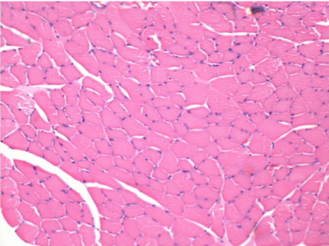
L : Control-100xD10
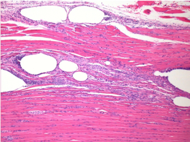
R:100xD10
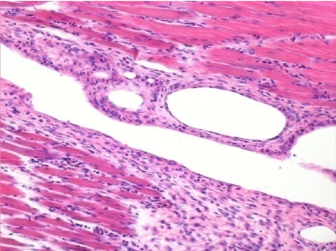
R :200xD10
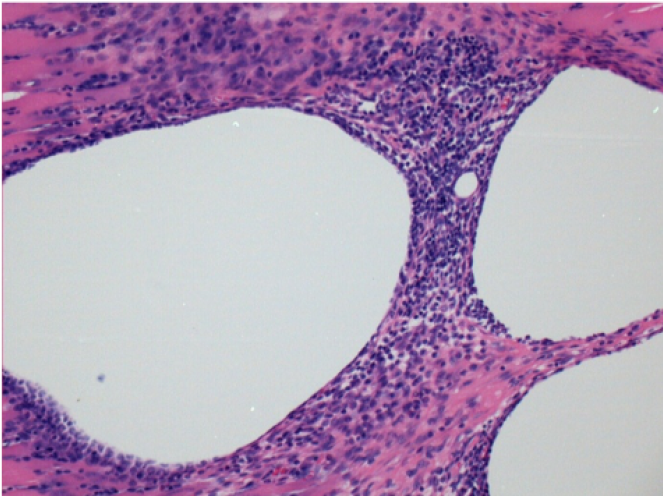
R :400xD10
07
1 month after Endopeel Injection
1 month after Endopeel Injection 0.1ml in the right pretibial muscle.
What is seen in black on the pictures is not a necrosis like could imagine some scientifics !
In fact, 4 conclusions have to be taken in consideration
- an artefact of coloration
- an absence of necrosis
- an apoptosis
- a bioregenerative process
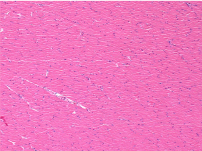
L : Control-100xD30
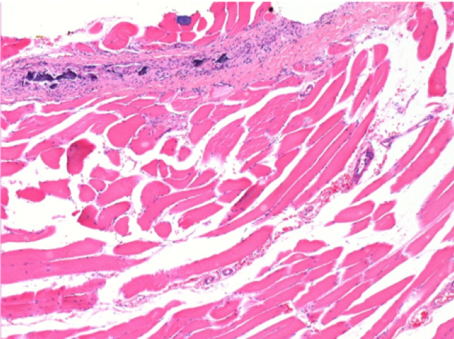
R:100xD30
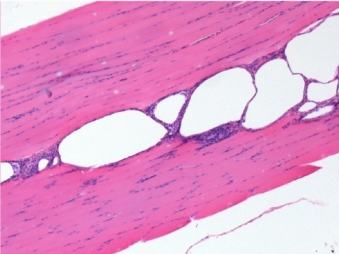
R :400xD30
08
3 months after Endopeel Injection
3 months (D90)after Endopeel Injection 0.1ml in the right pretibial muscle.
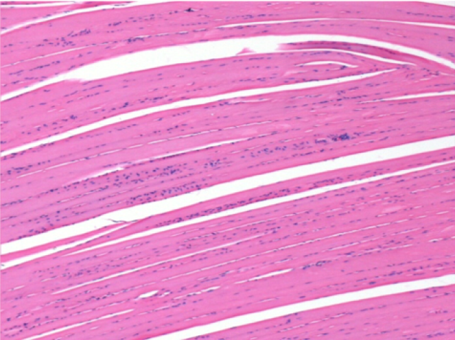
L : Control-100xD90
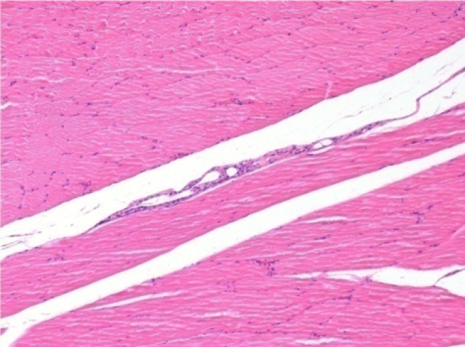
R:100xD90
09
7 months after Endopeel Injection
7 months (D210)after Endopeel IM Injection 0.1ml in the right pretibial muscle.
Complete Restitutio ad integrum after 7 months
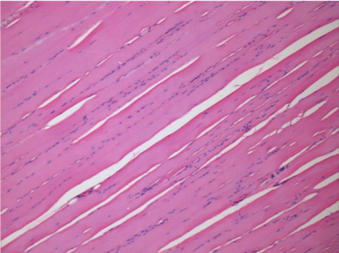
L : Control-100xD210
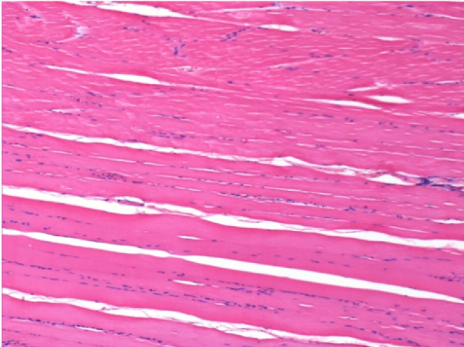
R:100xD210
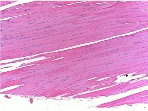
L :Control 50xD210
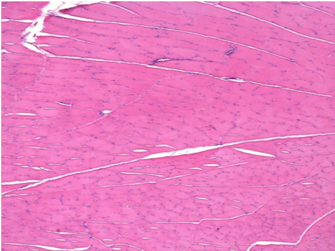
R50X-D210
10
Endopeel Injection in Subcutaneous Tissue
0.5 ml ( 5x 0.1ml) Endopeel SC Injection in the right subcutaneous pretibial area.
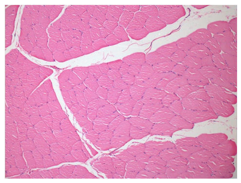
L:200x-Control-SC
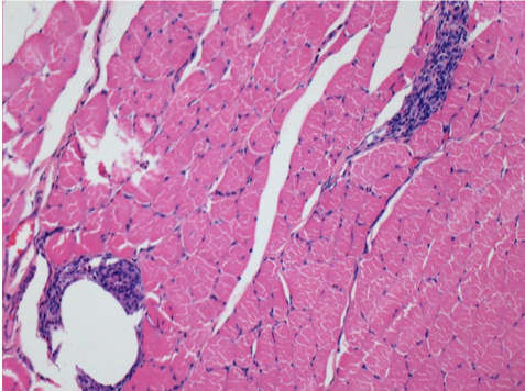
R-D10-SC-200X
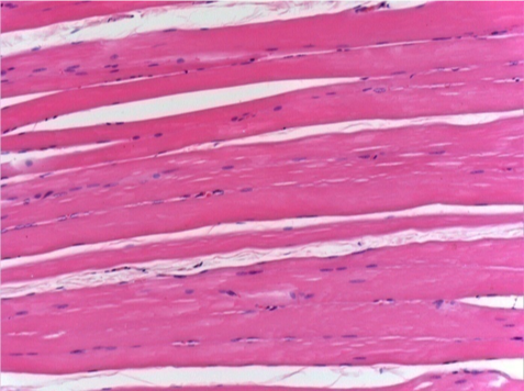
R-D30-SC-200X
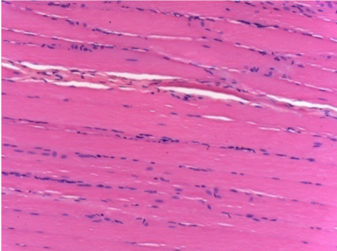
R-D90-SC-200X
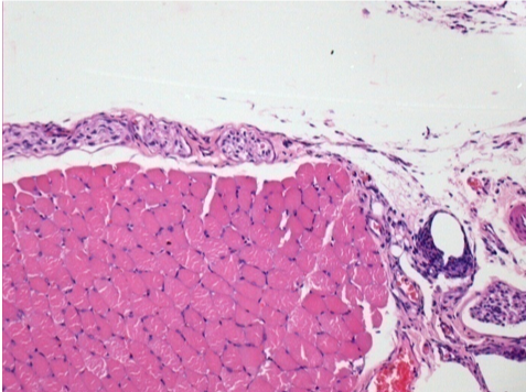
R-D210-SC-200X
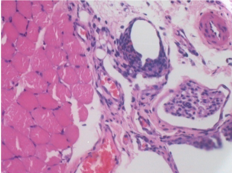
R-D210-SC-400X

-
- Endopeel induces a selective reversible myofibrolysis and inflammatory reaction on a period of 1 month, approximately
-
- Muscular changes are reversible in almost full totality
-
- The muscle is the better place to inject Endopeel because of more efficacity, control and duration of its action
-
- No necrosis nor abcess have been found all over the study.

